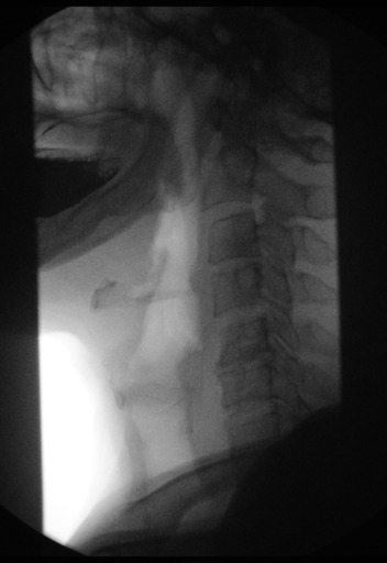
The whole goal of interventional radiology (IR) is to perform image-guided minimally invasive procedures on patients to both treat and diagnose conditions in nearly every organ of the body (so the IR department regularly coordinates with almost every division in the hospital). In most procedures, an interventional radiologist will gain access to a patient's circulatory system via the femoral artery by using an ultrasound device to find insert a catheter (small tube) into the artery. Once in the femoral artery, the radiologist will use a medical-grade wire guided by images from a machine called a "C Arm" to gain access to any part of the body. A C Arm produces continuous real time images of the wire going through the arteries. Since x-rays are used during these procedures, everyone in the operating room is required to wear lead vests and dosimeters (badges that measure cumulative exposure to radiation) at ALL times. Once the wire is in place, a microcatheter (very small tube) is put over the wire and inserted into the body. Using the mircocatheter, intravenous fluoroscopy (which uses contrast agents to highlight parts of the images) can be performed to highlight arteries on the x-ray.
 |
| Swallowing Contrast Agent (A form of Fluoroscopy) |
| C Arm |
The benefits of interventional radiology are evident in the procedures themselves. During my time in the department, general anesthetics were rarely used in any procedure. The incision is so minuscule and the procedures are so noninvasive that the patients are conscious for almost every part of the procedure (unless the sedatives force the patients to sleep.) One patient was completely lucid and even had a full conversation with the attending doctor and resident during a procedure. After procedures are over, almost every patient (the IR department works with "in" patients as well) leaves the hospital the very same day.
Despite how harmless these procedures may seem, they are extremely effective and are even preferential to traditional surgery in some cases. Interventional radiology is not a preferential treatment plan for patients that are in need of immediate care (i.e. unstable trauma victims), but is preferential for patients that are stable or bad candidates for surgery (i.e. patients that are older or have lost a lot of blood.)
During my time in IR, the most popular procedure was a port-a-catheter. The point of this procedure is to insert a port (which can be injected with medicine) under the skin and attach the port to a catheter that feeds into the end of the superior vena cava and directly into the heart. This procedure is for patients that are going to be regularly receiving medicine in quantities that veins could not handle (i.e. chemotherapy.) The heart is much stronger than the veins, so the heart can receive much stronger medication, as well as effectively distribute the medication to all parts of the body.
 |
| Port-a-Catheter |
The most memorable procedure I observed during my time in IR was on a patient that had been the victim of a trauma and was brought to interventional radiology by the trauma unit. The trauma unit had stabilized all of the patients injuries besides one - the patient's spleen was bleeding. The patient had already lost a substantial amount of blood and was not a good candidate for the surgical unit to remove the patient's bleeding spleen to stabilize the patient. The patient was brought to the IR operating room while still receiving a blood transfusion to keep the patient's blood pressure stable. The trauma surgeons were observing the procedure in hopes that the bleeding was not extensive and repairable. In order to determine how badly the spleen was bleeding, the IR doctors performed an angiogram of the spleen to find the origin of the bleeding, as well as the extent of the damage to the patient's spleen. Fortunately, the spleen did not need to be embolized to stop the bleeding. Instead, the IR doctors were able to use a type of foam that clots around damaged areas and stops bleeding. So in the end, the IR doctors saved the patient's spleen and the patient did not have to undergo open surgery to remove the spleen.
The week in IR kept me on my toes and I learned a lot. Interventional radiology gave me the opportunity to gather valuable information, as well as opened my eyes to many different types of medicine that I never knew were possible. Interventional radiology is a powerful tool that is constantly developing and refining treatments to effectively treat conditions with less of a risk to patients.
If I had a choice between surgery and IR, I would definitely choose IR.